Superconducting scanning tunneling microscope tip to reveal sub-millielectronvolt magnetic energy variations on surfaces
A superconducting tip yields the energy resolution to detect small shifts of spin-flip excitation thresholds, permitting the authors to reveal the different individual environments of molecules in an ordered layer. This knowledge allows Mier et al. to reveal the adsorption configuration of a complex molecular structure formed by molecules in different orientations and positions.
At low electron energies (i.e. small bias between a scanning tunneling microscope tip and the sample), the conductance between two superconductors shows enhanced peaks at a bias matching the sum of the sample and tip superconducting gaps. For the present experiment, the authors use a Pb (111) substrate and a nickelocene molecule. The tip is also covered by Pb, leading to the same superconducting gap for tip and substrate. The attached figure shows the corresponding peaks at 2Δ. Between 4 and 5 Δ a new large peak appears. This peak is due to an inelastic spin flip of the nickelocene molecule used in these experiments. The injected electron loses the energy needed to excite the molecular spin and ends up in the large density of states corresponding to the peaks at 2Δ. This leads to an enhanced inelastic peak at 2Δ plus the spin excitation energy. The resolution of the experiment is very good and permits to reveal a sub-milli electronvolt variation of the spin excitation. This spin excitation variation is traced back to the effect of the substrate on the molecule. Mapping the variation of excitation energy over the molecular overlayer on the surface allows the team to find the adsorption configuration of the molecule on the surface. The results are plotted on the right panel. They find an intricate geometry of molecular configurations with a non-commensurate registry with the surface.
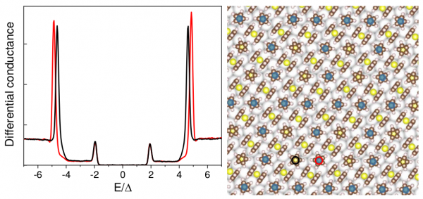
Figure: Differential conductance between a Pb tip and a Pb(111) substrate covered with nickelocene (C10H10Ni) molecules.
The molecule has a sandwich structure with two cyclopentadienyls (C5H5) above and below a Ni atom. Consequently, the molecule can lie flat or vertical on the surface (right panel). Moreover, the structure is not commensurate due to the very small local interactions between molecule and substrate. This leads to a different crystal field on the molecule depending on the adsorption site that reflects in a small (sub-milli eV) shift in the molecular magnetic anisotropy as revealed in the left panel. The shift is only present in the bigger-energy peaks, as it corresponds to the spin-flip origin of this peak. The lower-energy peaks correspond to the quasiparticle structure of tip and substrate and hence do not depend on the molecular adsorption.