Mapping Lamb, Stark, and Purcell Effects at a Chromophore-Picocavity Junction with Hyper-Resolved Fluorescence Microscopy
The properties of molecular transitions depend on their optical interaction with the surrounding environment. Confining light to atomic-sized hot spots pushes this effect to its ultimate limit, enabling the characterization and control of the energy and losses of a molecular transition with submolecular resolution.
The interaction of photons with excitonic and vibrational molecular transitions is often the target of state-of-the-art spectroscopic and microscopic techniques. This interaction can be enhanced by using optical nanoresonators that confine electromagnetic energy into tiny volumes of space. Plasmonic modes in metallic nanocavities are particularly attractive in this context, since they push light confinement to the limit as compared to standard dielectric resonators.
Confinement of photons in nanocavities enables to obtain optical images of molecular excited states with improved resolution. The characterization of different regions of the excitations in individual molecules (submolecular resolutions), is, however, a formidable challenge, only recently surmounted with the use of plasmonic picocavities in Scanning Tunnelling Microscopy (STM) configurations. Picocavities are created by atomistic features in an STM tip, strongly boosting electromagnetic fields in atomic-sized regions.
A theoretical and experimental study developed in a collaboration by the Theory of Nanophotonics group in CFM, the tunnelling microscopy group at the University of Strasbourg, and researchers at University Paris-Saclay, has demonstrated and interpreted submolecular resolution with the use of a picocavity not only in the optical characterization, but also in the manipulation of the properties of excitonic molecular transitions. As the tip of a STM is placed over different positions on top of a phthalocyanine molecule, a strong change of the energy and decay rate (effective losses) of an excitonic transition of the molecule is measured. The increase of the decay rate is produced as a consequence of the optical coupling between the plasmonic picocavity and the molecule (Purcell effect). Understanding the changes of the transition energy induced by the tip position requires to consider both the electrodynamical coupling (Lamb shift) as well as the electrostatic interaction (Stark shift). This work thus emphasizes the exquisite control of the optical properties of single molecules achievable in STM, as well as the subtle origin of some of the spectroscopic and microscopic features.
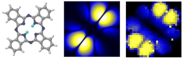
Figure: Left- Experimental map of light emission from a free-base phthalocyanine (H2Pc) deposited on an NaCl-Ag(111) surface when scanned in a STM picocavity. The area of scanning is 2.5 x 2.5 nm2, and the bias voltage applied is V=-2.5 V, with a current I= 100 pA. Right- Theoretical map of light emission under the same circumstances as in the experiment. The spectral range of light emission considered is at the excitonic emission line 1.975 eV.