Vibrational Electron Energy Loss Spectroscopy in Electron Microscopy reveals new aspects of water at the nanoscale level
A new microscopy technique developed at the University of Illinois at Chicago, in collaboration with researchers at Oak Ridge National Lab. in Tennessee, the Nion Company in Seattle, and the Center for Materials Physics in San Sebastian, Spain (CSIC-UPV/EHU) allows researchers to visualize liquids at the nanoscale level – about 10 times more resolution than with traditional transmission electron microscopy – for the first time, and to understand the details of the vibrational properties of the liquid sample in a complex nanoenvironment.
By trapping minute amounts of liquid between two two-dimensional layers of boron nitride, the liquid sample can be imaged at extremely high resolution using a traditional transmission electron microscopy and spectroscopy techniques. This approach could provide information on the vibrational state of individual molecules.
The new technique can be used to follow nanoscale-sized tracers used in biological research, and to visualize processes at liquid-solid interfaces at unprecedented resolution. Using their specialized sample holder, or boron nitride cell, the researchers describe the unique vibrational properties of water and heavy water at the nanoscale level. Researchers of the “Theory of nanophotonics” group in San Sebastian helped to interpret the vibrational information obtained in the energy loss spectra, by a modelization of infrared response of the liquid water sandwiched in the boron nitride layers. This study shows the importance of the nanoscale environment to properly account of the energy and intensity of the vibrational lines obtained with this novel microscopy technique.
The experimental researchers at Chicago and Tennessee had to solve the problem of how to isolate tiny amounts of liquid in preparation for scanning transmission electron microscopy, which uses a focused beam of electrons to image samples. Normally, samples must be frozen or encased in epoxy and then sliced superthin before being placed under the electron beam, where the user has just seconds to take pictures of the sample before it vaporizes. After dismissing other options, the researchers finally settled on nanolayers of boron nitride to encapsulate and support the liquid without affecting the measurements.
By using a scanning transmission electron microscope with one of the world’s best energy resolutions at the Department of Energy’s Oak Ridge national laboratory in Tennessee, the vibrational spectrum of liquid water at the mili-electron volt’s range could be measured. Normally, water in large amounts vibrates at 420 mili-electron volts, but the researchers witnessed that water trapped in the BN cell vibrated at 406 mili-electron volts.
This new electron microscopy technique allows to see physical and chemical processes happening in a liquid environment at the nanoscale level. Far smaller volumes can be measured by other methods, but at such small scales, the behavior of water changes as individual atomic bonds, local electric fields and the proximity of surfaces begin to affect its normal behavior. Some of these effects have been accounted for within the dielectric response theory used by the “Nanophotonics Theory” group at the Center for Materials Physics in San Sebastian to reveal the complex interactions of the nanoenvironment which determines the final details of the vibrational spectra.
This work has been published in Advance Materials, and has been funded in part by National Science Foundation grants DMR-0959470 and DMR-1626065 and by grant FIS2016-80174-P from the Spanish Ministry.
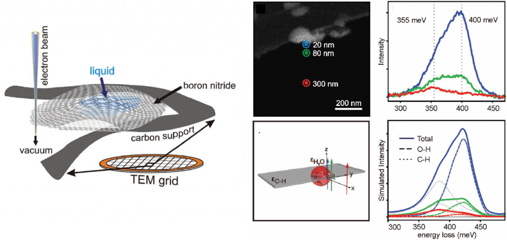
Figure: Left: Schematics of the sample consisting of liquid water encapsulated in a double layer of boron nitride supported on carbon at the TEM grid of the scanning transmission electron microscope. Right: Top-left: Image of the cell probed by electron beams at three different positions (blue, green, and red). Top-right: Experimental vibrational electron energy loss spectroscopy (EELS) of the sample to the left for the three different electron beam positions. Bottom-left: Model system used to calculate the dielectric response of a water bubble supported by a carbon layer. Bottom-right: Theoretical vibrational EELS of the model system to the left reproducing the vibrational fingerprints of the liquid nanoscale system, revealing the O-H and C-H stretching modes.