An international team, with the participation of the Donostia International Physics Center (DIPC) and the Centre of Materials Physics (CSIC-University of the Basque Country UPV/EHU), has resolved and identified, with a hitherto unprecedented resolution, a single organic molecule using light. The prestigious journal Nature has just published the research work, which opens doors to possible technological applications with biosensors.

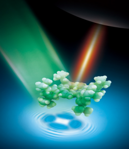
Artist’s view of a light beam (green) illuminating a plasmonic cavity, which enhances the electromagnetic field at the cavity (red light). Thanks to this antenna effect, the vibrational map of a single molecule and its inner structure (shadow below the molecule) can be resolved and identified with unprecedented resolution with visible light.
A research work led by Prof. Zhenchao Dong at the University of Science and Technology of China, in Hefei, and in which Javier Aizpurua, head of the “Theory of Nanophotonics Group” at the DIPC and the CSIC-UPV/EHU has participated, has managed to resolve and identify for the first time a single organic molecule with a subnanometric-range resolution, using light. In the words of Javier Aizpurua: “We have been able to see <<inside>> a single molecule and identify what kind of molecule it is, simply by using light”. The results have been published in the last issue of the prestigious journal Nature.
Visible light is an electromagnetic wave whose wavelength is between 400 nanometres (nm), (blue) and 750 nm (red). Due to what is known as the resolution diffraction limit, using light, it is impossible to directly resolve or photograph objects with a size less than half of the wavelength of the light, i.e. less than 200 nm. In order to overcome this limitation, over recent years specialists in Nanophotonics have used metal particles that act as minute optical antennae, concentrating and enhancing the visible light spectrum on a nanometric scale. However, even this technique has its limitations and difficulties when trying to resolve nanometric objects.
The optical resolution achieved in this research, hitherto never obtained, has been possible thanks to the combined use of the scanning tunneling microscope (STM) technique in ultra-high vacuum and low temperature conditions, with the Tip-enhanced Raman spectroscopy (TERS) technique, which dramatically enhances the field that acts on the molecule located in the cavity of the tip of the microscope.

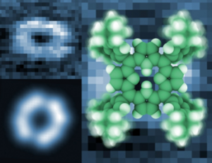
Top left: experimental map of an isolated porphyrin molecule for a given vibration frequency. Bottom leff: theoretical calculation of the same molecular vibration showing its fingerprint. On the right: molecular structure of the porphyrin used in the experiment.
The combination of these two techniques has enabled “photographing” organic molecules for the first time at a subnanometric scale. The tuning of the collective oscillation of conduction electrons at the tip of the microscope, the so-called plasmons, with the vibrational excitation of the molecule, enables generating a non linear optical signal with a sub-nanometric resolution.
When the microscope tip is scanned over the molecule, the Raman signal emitted at each point enables identifying the vibrational signature of the molecule in such a way that, apart from looking “inside” the molecule, it is simultaneously possible to identify which molecule is involved. Javier Aizpurua explained that “it is like peering <<inside>> the molecule and taking its fingerprints”.
This level of resolution has only been possible to date using electrons as the probes, but in this research it is the photons of visible light that manage to achieve the miracle of identifying a molecule, going beyond all limits of optical diffraction until now known.
The results of this work open the doors to the direct identification of molecules when their concentration is very small, managing to identify even a single isolated molecule. This ability gives rise to a wide range of possible technological applications, such as in biosensor ones for the analysis of molecular chains, in health and safety for detecting dangerous substances, and in public health for the control of food quality, amongst others.
Original Publication: Chemical mapping of a single molecule with sub-nm resolution by plasmon-enhanced Raman scattering. R. Zhang, Y. Zhang, Z. C. Dong, S. Jiang, C. Zhang, L. G. Chen, L. Zhang, Y. Liao, J. Aizpurua, Y. Luo, J. L. Yang, and J. G. Hou. Nature DOI: 10.1038/nature12151

